Abstract
Introduction: Gaucher Disease (GD) is a rare disease caused by recessive mutations in the glucocerebrosidase A gene (GBA1) that cause a deficiency in acid-glucocerebrosidase enzyme at lysosomes and the accumulation of glucosylceramide. Haematological involvement is identified as bone marrow (BM) infiltration by Gaucher cells, thrombocytopenia and anemia; the bone involvement is registered as bone pain, vascular complications, deformation and aseptic osteomyelitis. Toghether they are the main symptomatology in the type-1 or non-neuronopatic GD. The bone affectation is the leading cause of morbidity and disability in GD patients. Current available therapies decrease the bone marrow infiltration and the incidence of bone complications. However, the identification of risk-factors to predict bone complications (that can occur even in patients under long-term ERT) or guide the therapy are in need.
Aims: In this work a combination of a structured-MRI report for GD an a machine learning methodology has been applied in order to identify features to predict GD bone complications.
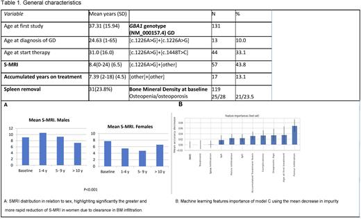
Patient and Methods: Retrospective descriptive study using the data base of the Fundacion Espanola para el Estudio y Terapeutica de la Enfermedad de Gaucher (FEETEG). General, genetic and clinical data from GD patients who has been evaluated in the FEETEG unit with MRI were included. All the MRI assessments were re-evaluated in a blinded manner for an expert radiologist in order to generate an structured MRI BM report registering the infiltration in the lumbar spine, pelvis and femora according to the Spanish MRI score (S-MRI) from Roca et al. assigning 4-points for homogeneous pattern, 3-points for non-homogeneous diffuse, 2-points for non-homogeneous mottled and 1-point for reticular pattern for each region. The presence of bone complications (infarcts, necrosis, fractures, arthropathy or bone crisis) added 4 points for each complication observed in the MRI. The MRI reports were grouped in 1-at diagnosis, 2-between 1-4 years of follow-up (FU), 3-from 5-9 years of FU and 4-after 10+ years of FU. Three Random Forest models were designed and trained to identify features to predict bone disease progression and complications; model A included all of the variables chosen for the Machine Learning study, model B considered whether a treatment was applied or not, ignoring the particular treatment, and model C ignored the S-MRI score. The model parameters were optimized using a grid search. Model performance on the validation dataset was evaluated using the ROC curve, AUC, accuracy and f1-score. The model was created using the scikit-learn package for python 3.10.4. The study was approved by the FEETEG ethic and scientific boards.
Results: Data from a total of 131 patients, 62 females with a total of 441 MRIs, 131 at first visit and 310 at FU were included. See table 1 for general characteristics. At first MRI, the mean S-MRI score was 8.4 (95% CI 0-25), with males showing a higher score (9.1) compared to females (7.71) (p<0.001); also, 63 patients (48.4%) showed vascular complications at the first MRI. The heterozygosity for c.1226A>G/other or other/other were associated with differences in BM infiltration when compared with c.1226A>G homozygosis or c.1226A>G /c.1448T>C cases (p=0.017). Splenectomy was related with high S-MRI score (p<0.001) and higher incidence of bone complications (p<0.001) as expected. In the FU MRI, females showed a faster reduction of BM infiltration (see Fig 1a). 61.5% (80) of patients showed infiltration in spine, pelvis and femur at baseline. A homogeneous MRI pattern was reported in 36.1% in lumbar spine, 10.7% in pelvis and 3.8% in femurs. The three random Forest model achieved AUC of 75.82%, 85.73% and 69.76% (accuracies 78.1%, 83.81% and 74.29% respectively), all with a decision boundary threshold of 0.5. The most important features identified were: S-MRI score, age of first treatment and treatment for model A; S-MRI score, age of first treatment and spine infiltration for model-B and the femur infiltration, age of first treatment and age at diagnosis for model-C.
Conclusions: In the studied population, men showed a higher level of infiltrations and women a lower and faster response to therapy. The random forest model identified for the firs-time the presence of BM infiltration at femur level and the presence of a homozygous infiltration pattern at any level as predictors for bone complication
Disclosures
Giraldo:Takeda: Consultancy, Honoraria; Acindes: Consultancy, Honoraria; Alexion: Consultancy, Honoraria, Membership on an entity's Board of Directors or advisory committees; ZeroLMC Foundation: Honoraria, Membership on an entity's Board of Directors or advisory committees; Sanofi: Consultancy, Honoraria.
Author notes
Asterisk with author names denotes non-ASH members.


This feature is available to Subscribers Only
Sign In or Create an Account Close Modal